Clinical features of patients presenting with choroidal melanoma at Eye Foundation Hospital Ikeja, Lagos, Nigeria
Abstract
BACKGROUND
Ocular melanomas are the most common primary intraocular tumors in adults, with the choroid being the most frequently affected uveal site.1,2 Their incidence is highest among populations of European ancestry and lowest among Asian and African populations.1,2 Clinical presentation varies depending on tumor location. Diagnosis is typically non-invasive, relying on slit-lamp examination, fundus evaluation, and ultrasonography.3 This case series describes four patients seen between 2010 and 2025 with choroidal melanomas, highlighting their clinical presentations, fundus findings, and ultrasonographic features (A- and B-scan).
CASE SERIES
Case 1
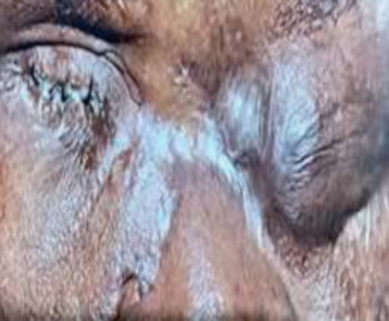
A 58-year-old female of British (Caucasian) origin presented with a 6-week history of progressive vision loss in the right eye. Visual acuity (VA) was 3/60 (right) and 6/6 (left). Fundoscopy revealed a large pigmented mushroom-shaped lesion in the inferotemporal quadrant, within 2 disc diameters of the optic disc, measuring approximately 8 disc diameters, with adjacent subretinal fluid. Cup–disc ratio was 0.2. The left fundus was normal. Ultrasound B scan showed an inferotemporal dome-shaped mass with low reflectivity on A-scan and orbital shadowing.(Table 1, Figure 1a) A diagnosis of right choroidal melanoma was considered, and patient was referred to an ocular oncology center in her home country.
Case 2
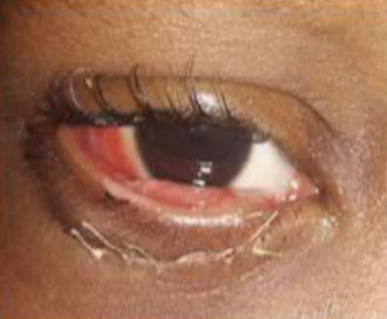
A 46-year-old male of African descent presented with complaints of an overhanging shadow in the superior visual field of the right eye. VA was 6/6 bilaterally. Fundoscopy showed a dome-shaped hyperpigmented inferonasal mass overlapping the optic disc, measuring 8 disc diameters, with perilesional orange pigments but no sentinel vessels or subretinal fluid. The left fundus was normal. Ultrasound revealed a mushroom-shaped mass with homogeneous opacity on B-scan, low internal reflectivity on A-scan, choroidal excavation, and orbital shadowing.(Table 1, Figure 1b) A diagnosis of right choroidal melanoma was made, and the patient was referred to the oncology unit of a tertiary hospital.
Case 3
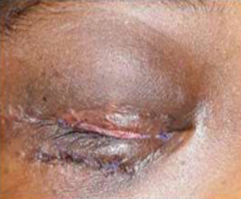
A 45-year-old female of African descent presented with reduced vision in her left eye. VA was 6/6 (right) and 6/18 (left). Fundoscopy showed a large parapapillary hyperpigmented mass (>12 disc diameters) in the nasal and superonasal quadrants, with indistinct margins and extensive subretinal hemorrhage obscuring the optic disc.
Ultrasound demonstrated a large dome-shaped choroidal mass with low reflectivity, choroidal excavation, orbital shadowing, and associated exudative retinal detachment.(Table 1, Figure 1c) A diagnosis of left choroidal melanoma was considered, and the patient was referred to the teaching hospital’s ocular oncology service.
Case 4
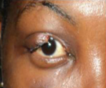
A 29-year-old male of African descent presented with a 1-month history of progressive visual loss, diplopia, and floaters in the right eye. He had previously received 3 intravitreal bevacizumab injections at another facility without improvement. His initial diagnosis was not available for review. VA was 6/12 (right) and 6/6 (left). Fundoscopy showed a large superotemporal choroidal mass overlapping the optic disc (~10 disc diameters), with associated exudate and exudative retinal detachment. The left eye was normal.
Ultrasound revealed a large dome-shaped choroidal mass with exudative detachment. A-scan showed low internal reflectivity with choroidal excavation and orbital shadowing.(Table 1, Figure 1d) Right choroidal melanoma was diagnosed, and a Positron Emission Tomography (PET) scan was requested, but the patient was lost to follow-up.
DISCUSSION
This case series highlights the rarity of choroidal melanoma in our setting: only four patients were diagnosed over 15 years (2010–2025). This reflects the lower incidence of ocular melanoma in African populations, possibly due to the protective effect of increased choroidal melanin in darker skin.4,5 Ultrasonography was the principal diagnostic tool, with consistent findings across cases: dome- or mushroom-shaped masses, low internal reflectivity on A-scan, homogeneous opacities on B-scan, choroidal excavation, orbital shadowing, and frequent association with subretinal fluid or exudative retinal detachment.6,7 Outcomes of management of reported cases were not available to the authors.The low awareness of intraocular tumors in Africa may delay diagnosis.8,9 Increased clinical vigilance is therefore warranted, even in populations with low incidence.
References
1. Liang X, Liu W. Characteristics and Risk Factors for Spontaneous Closure of Idiopathic Full-Thickness Macular Hole. J Ophthalmol. 2019 Mar 13;2019:1–6.
2. Patel DR, Blair K, Patel BC. Ocular Melanoma. [Updated 2024 May 2]. In: StatPearls [Internet]. Treasure Island (FL): StatPearls Publishing; 2025 Jan-. Available from: https://www.ncbi.nlm.nih.gov/books/NBK551647.
3. Soliman N, Mamdouh D, Elkordi A. Choroidal Melanoma: A Mini Review. Medicines. 2023 Jan 5;10(1):11.
4. Miller B, Abrahams C, Cole GC, Proctor NS. Ocular malignant melanoma in South African blacks. Br J Ophthalmol. 1981 Oct;65(10):720–2.
5. Singh P, Singh A. Choroidal melanoma. Oman J Ophthalmol. 2012;5(1):3.
6. Sabazade S, Herrspiegel C, Gill V, Stålhammar G. No differences in the long-term prognosis of iris and choroidal melanomas when adjusting for tumor thickness and diameter. BMC Cancer. 2021 Nov 24;21(1):1270.
7. Ramos-Dávila EM, Dalvin LA. Clinical Implications of Ultrasound-Based Morphology in Choroidal Melanoma. Ophthalmol Retina. 2025 Mar;9(3):263–71.
8. Lukamba RM, Yao JA, Kabesha TA, Budiongo AN, Monga BB, Mwembo AT, Bey P, Chenge GB, Desjardins L, Luboya ON, Doz F, Stefan CD. Retinoblastoma in Sub-Saharan Africa: Case Studies of the Republic of Côte d'Ivoire and the Democratic Republic of the Congo. J Glob Oncol. 2018 Sep;4:1-8. doi: 10.1200/JGO.17.00056. PMID: 30241234; PMCID: PMC6223407.
9. Ayanniyi AA, Jamda AM, Badmos KB, Adelaiye RS, Mahmoud AO, Kyari F, Nwana EJ. Awareness and knowledge of ocular cancers in a resource-limited economy. N Am J Med Sci. 2010 Nov;2(11):526-31. doi: 10.4297/najms.2010.2526. PMID: 22558560; PMCID: PMC3338215.
Additional Files
Published
How to Cite
Issue
Section
Categories
License
Copyright (c) 2025 Transactions of the Ophthalmological Society of Nigeria

This work is licensed under a Creative Commons Attribution-NonCommercial-NoDerivatives 4.0 International License.